Denver’s Trusted Chiropractor for Lasting Pain Relief, Not Just Temporary Fixes
What’s Covered on This Page
- What Advanced X-Ray Analysis Actually Measures
- Why Weight-Bearing Positioning Changes the Diagnosis
- Conditions That Require Imaging Before a Chiropractic Adjustment
- What to Expect at Your Advanced X-Ray Analysis Appointment
- How X-Ray Findings Directly Shape Each Chiropractic Adjustment
- Do I need to do anything to prepare before my advanced X-ray analysis appointment in Denver?
- What happens after my X-rays are taken — how soon will I know what’s going on?
- I’ve already had X-rays somewhere else and was told everything looks fine — why would yours show something different?
- Is it safe to get X-rays at a chiropractic office, and how much radiation is involved?
- Denver has a lot of desk workers and remote employees — does that affect what you typically find on X-rays?
- Can you do advanced X-ray analysis on kids or teenagers, or is it just for adults?
What Advanced X-Ray Analysis Actually Measures
Most people think an X-ray just shows bones. Broken or not broken. That’s about it, right? But advanced X-ray analysis goes deeper than that, it measures the actual geometry of your spine.
We’re looking at angles. Curves. Alignment from one vertebra to the next. Every degree matters because your spine has very specific measurements it should fall within. When those numbers are off, your body compensates. And that compensation is usually what’s causing your pain.
Here’s what we measure on every set of films we analyze:
- Cervical lordosis angle, which tells us if your neck curve has straightened or reversed
- Lumbar lordosis, the natural low back curve that absorbs shock when you walk or sit
- Disc space height between vertebrae, showing early degeneration before you feel it
- Vertebral rotation and lateral shift, common in patients with scoliosis or chronic one-sided pain
- Pelvic tilt and sacral base angle, which affect everything above them
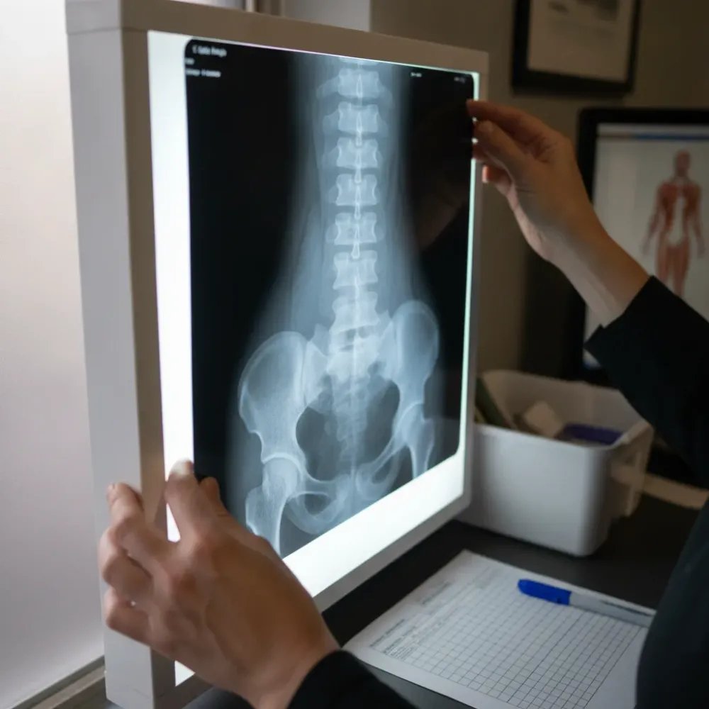
These aren’t guesses. We draw lines on your films. We calculate angles down to the degree. Patients who’ve been told “your X-ray looks fine” somewhere else come to us and we find measurable structural problems that were simply overlooked.
So why does this matter to you? A 12-degree loss in your neck curve creates real mechanical stress. Your head weighs about 10 to 12 pounds. When that curve flattens, the muscles in your neck and upper back work overtime just to hold your head up. Patients across Denver sit at desks all day and don’t realize their neck curve has been slowly disappearing for years.
A standard X-ray gets read for fractures and obvious pathology. Advanced X-ray analysis gets read for function. We want to know how your spine is performing under gravity, not just whether something is visibly broken. That’s the difference between managing symptoms and building a correction plan that changes your structure over time.
Why Weight-Bearing Positioning Changes the Diagnosis
Most X-rays you’ve had were probably taken lying down. That’s standard at hospitals and urgent care clinics. But here’s the problem. Your spine doesn’t work lying down. It works standing up, carrying your body weight, fighting gravity every second of the day.
When we refer patients for imaging, we specify weight-bearing positioning whenever clinically appropriate. That means you’ll be standing during the films, not lying flat on a table. Most imaging centers can accommodate this when the order specifies it. The difference in what shows up is significant.
We see this all the time in our Denver office. A patient brings in X-rays from a previous provider, everything looks “normal.” They’re frustrated because they know something is wrong. Their back hurts. Their neck is stiff. Nobody can tell them why.
Then we take weight-bearing films. Standing up. And suddenly the picture changes completely.
When you’re lying flat on a table, gravity isn’t compressing your spine. Disc spaces look fine. Alignment looks acceptable. But stand that same person up and you’ll often see things that were invisible before:
- Disc spaces that narrow under load, revealing early degeneration
- Spinal curves that shift or worsen with gravity pulling on them
- Pelvic imbalances that only show when your full body weight is on your feet
- Vertebral misalignments that self-correct when you lie down but return the moment you stand
This is why someone’s been told “your X-rays look fine” even though they’re in real pain. The images weren’t taken in the position where the problem actually shows up. Weight-bearing imaging gives a more accurate view of spinal biomechanics than lying flat, and that difference matters when we’re building your care plan.
Think about it this way. You wouldn’t test a bridge by laying it flat on the ground. You’d test it under stress, with weight on it. Your spine deserves the same honest look.
For many patients, this is their first experience with advanced X-ray analysis done in a weight-bearing position. The difference in what we can see is dramatic and changes the entire treatment plan. A wrong diagnosis leads to wrong treatment. We’d rather get it right from the start.
Conditions That Require Imaging Before a Chiropractic Adjustment
You’d be surprised how many people walk into our Denver office thinking their back pain is “just a muscle thing.” And sometimes it is. But sometimes there’s something deeper going on, something we need to see before we put our hands on your spine.
That’s the whole point. Not every patient needs X-rays. But certain conditions make imaging non-negotiable.
Here are situations where we won’t adjust you without looking at your films first:
- History of a car accident or trauma. Even a minor collision can shift vertebrae in ways you won’t feel for weeks. We need to rule out fractures, ligament damage, and instability before any spine adjustment happens.
- Scoliosis or suspected spinal curvature. If one shoulder sits higher than the other or your hips are uneven, we want to measure exactly what’s happening. Adjusting a curved spine without imaging is guessing.
- Pain that hasn’t responded to other treatments. You’ve tried stretching, rest, maybe even another chiropractor. Nothing worked. That’s a sign something structural is being missed.
- Numbness, tingling, or radiating pain. These symptoms often point to a pinched nerve or disc issue. We see this every week, especially in patients dealing with sciatica pain.
- Osteoporosis or bone density concerns. Particularly common in our seniors. Bones that have lost density respond differently to force, so we need to know what we’re working with.
We also image patients who’ve had prior spinal surgery. Post-surgical spines have hardware, fusions, scar tissue. Imaging before treatment in these cases is considered standard of care. We take that seriously. The CDC provides a helpful radiation imaging procedures overview that outlines what patients should understand about diagnostic imaging and radiation safety before undergoing any X-ray procedure.
Here’s what I tell patients in our office. If you’ve been dealing with pain for more than a few weeks, or if something just feels wrong, imaging gives us both peace of mind. It takes the guesswork out completely. We’re not ordering films to run up a bill, we’re doing it because your safety matters more than speed.
Most people feel relieved once they can actually see what’s going on inside their own body. That clarity changes everything about how we build your care plan together.
Need help with Advanced X-Ray Analysis?
Schedule Your Initial Examination. Glendale Chiropractic is ready to help.
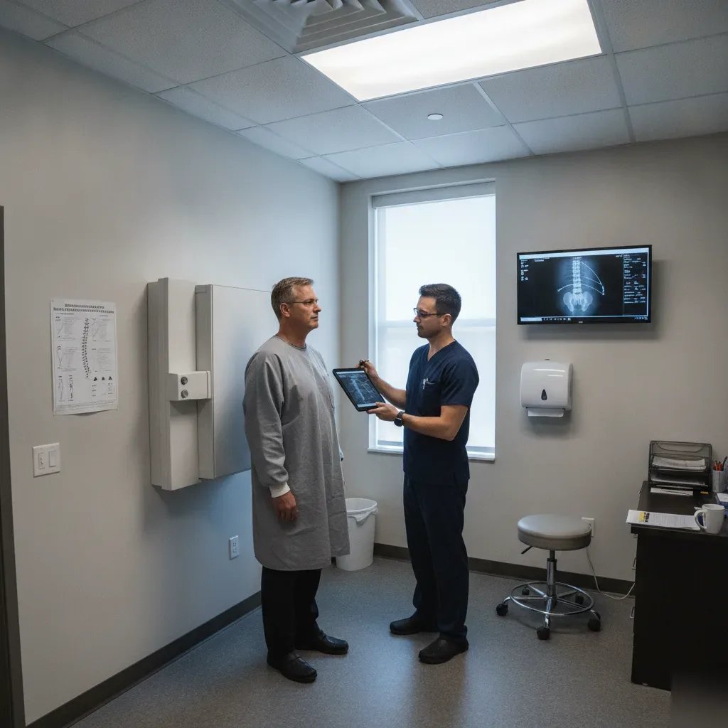
What to Expect at Your Advanced X-Ray Analysis Appointment
You walk in, we talk. That’s how it starts.
Before we take a single image, we sit down and go over what’s been bothering you. Where the pain shows up, how long it’s been there, what makes it worse. We want to hear your story because your symptoms tell us exactly where to look. Most people who come to our Denver office have already tried a few things that didn’t work. That frustration is real, we hear it every day.
Once what we’re dealing with, here’s how the appointment flows:
1) We refer you to an imaging center with a specific order detailing exactly which views we need and how you should be positioned.
2) Once your films come back, we perform the full analysis here in our office.
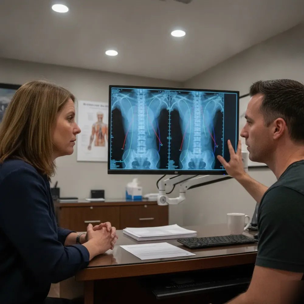
3) At your follow-up visit, we sit down with you and walk through every finding on screen. You’ll look at your own spine and actually understand what’s going on.
The whole process usually takes less time than people expect. And you won’t leave wondering what happens next.
Here’s what makes this different from the X-rays you’ve had at urgent care or a hospital. We’re not just looking for fractures or tumors. We’re measuring alignment, checking for structural shifts, and identifying patterns that explain why your pain keeps coming back. The answer is often sitting right there in the image.
Patients from across Denver have told us the same thing: “Nobody ever showed me my X-rays before.” We always do. When you can see the problem with your own eyes, the path forward makes sense. Dr. Brockway has over 15 years of experience reading spinal imaging and will explain every finding in plain language. No jargon. No guessing.
Ready to see what’s really going on? Give us a call.
How X-Ray Findings Directly Shape Each Chiropractic Adjustment
Here’s where advanced X-ray analysis stops being a diagnostic tool and starts being your treatment roadmap. Every adjustment we deliver in our Denver office is built on what we actually see in your images. Not guesswork. Not a standard protocol we run on every patient who walks through the door.
We see this every week. Someone comes in with neck pain and they’ve been adjusted the same way at three different offices. Nobody ever looked at the structure underneath. When we pull up their X-rays, we might find a reversed cervical curve or a vertebra rotated in a direction nobody accounted for. That changes everything about how we approach your spine.
Think about it this way. Your X-ray findings tell us three things that matter most:
- The exact angle and direction each misaligned vertebra needs to move
- How much force is safe to use based on bone density and disc spacing
- Which segments to leave alone entirely because they’re compensating, not causing the problem
That third one surprises people. Sometimes the spot that hurts isn’t the spot that needs correcting. Your body shifts and compensates to protect the real problem area. Without X-ray findings guiding us, a chiropractor might adjust the compensation pattern and actually make things worse.
So when we sit down with you and review your images, we’re mapping out a specific correction plan. This means your chiropractic adjustment isn’t generic. If your X-rays show a 12-degree lateral shift at L4, that tells us the precise vector for that spine adjustment. If there’s disc narrowing at C5-C6, we’ll use gentle chiropractic adjustments in that region instead of a traditional manual approach.
Most patients tell us nobody ever explained their X-rays to them before. That’s a problem we take seriously.
Imaging-guided care helps us identify contraindications and tailor treatment to your individual spinal anatomy. Your X-rays aren’t filed away after day one. We reference them throughout your care, adjusting our approach as your structure changes and improves over time. For cases where spinal instability or ligament damage is suspected, we may also order a motion x-ray analysis to capture how your spine behaves during movement. Need help figuring out what’s driving your pain? Give us a call.
Frequently Asked Questions
Common questions about Advanced X-Ray Analysis in Denver
Do I need to do anything to prepare before my advanced X-ray analysis appointment in Denver?
You don’t need much preparation before your visit. We refer you to an imaging center for the actual films. When you go, wear comfortable loose clothing and leave jewelry at home. The imaging center will position you standing for weight-bearing views when our order specifies it. That’s different from most clinics. Just show up ready to stand still for a few seconds per image. Bring any previous X-rays or MRI films you have, we review those too.
What happens after my X-rays are taken — how soon will I know what’s going on?
Once your films come back from the imaging center, we perform the full analysis here. At your follow-up visit we walk through every finding together on screen. We don’t just hand you a report and send you home. We draw the measurement lines on your films and walk you through each angle together. You’ll see exactly where your spine is out of range and why that matters for your pain. Most patients tell us this is the first time anyone actually explained their X-rays to them. From there, we build a care plan based on what the numbers show.
I’ve already had X-rays somewhere else and was told everything looks fine — why would yours show something different?
Most X-rays taken at urgent care or hospitals are read for fractures and obvious problems. They’re not analyzed for spinal geometry or biomechanics. We measure angles, curves, and alignment down to the degree. We also specify standing positioning when we refer patients for imaging. A spine that looks acceptable on a table can show real structural problems the moment gravity is involved. Many Denver patients come to us after being told their X-rays were normal, and we find measurable issues that were simply never looked for.
Is it safe to get X-rays at a chiropractic office, and how much radiation is involved?
Yes, chiropractic X-rays are safe and use a very low dose of radiation. Spinal films involve far less radiation than a CT scan. We follow standard safety protocols and only image when there’s a clinical reason to do so, we don’t X-ray every patient automatically. The CDC provides a radiation safety overview for patients who want to understand diagnostic imaging exposure before their visit. If you have concerns about radiation, just ask us directly, we’re happy to explain what we’re taking and why.
Denver has a lot of desk workers and remote employees — does that affect what you typically find on X-rays?
It absolutely does. We see this pattern constantly in Denver. Sitting for long hours, especially with a laptop or monitor at the wrong height, slowly flattens the natural curve in your neck. On X-rays, we measure this as a loss of cervical lordosis. A healthy neck has a 40-plus degree curve. Many desk workers we see are down to 20 degrees or less. That puts serious strain on the muscles holding your head up all day. Catching this early, before disc degeneration sets in, makes correction much more achievable.
Can you do advanced X-ray analysis on kids or teenagers, or is it just for adults?
Yes, we image younger patients when there’s a clinical reason, most often for scoliosis screening or after a sports injury. Scoliosis often shows up during growth spurts, and catching a curve early makes a real difference in outcomes. We use the lowest effective radiation settings for pediatric films. We won’t image a child without a clear reason to do so. If your teen has uneven shoulders, one hip higher than the other, or chronic back pain, that’s worth a conversation before assuming it’s just growing pains.
Ready to Get Started?
Schedule Your Initial Examination. Call +17208891659 today.